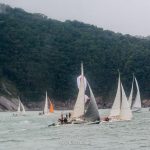
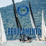
>) on both side of the image to move the images.>>) on both side of the image to move the images. We identified 1,002 tissue-specific genes that are a source of novel tissue-specific markers for 37 different anatomical structures." The nose is the body's primary organ of smell and also functions as part of the body's respiratory system. Morphological Mouse Phenotyping: Anatomy, Histology and Imaging is an atlas of explanatory diagrams and text that guides the reader through normal mouse anatomy, histology, and imaging. Latex injection apparatus. Histology atlas including Kaufman's Atlas of Mouse Development ... more info. Mouse embryo anatomy, ontology and staging ... more info. Comparative Anatomy of the Mouse and Rat: a Color Atlas and Text provides detailed comparative anatomical information for those who work with mice and rats in animal research. Rodent pancreas is soft and diffuse compared with the human pancreas. The probability maps for individual anatomical volumes were defined by the eight registered MR images used to construct the atlas, which serve as references to map the atlas onto test images and to label the regions of interest of test images. Given these differences between human and mouse glia, it is perhaps not surprising that where gene expression differs most between mouse and humans is in glia and not neurons. level of 3-D annotation for informatics analysis and viewing in the Brain Explorer® 3-D viewer. Take online anatomy classes taught by experts. Superficial muscles are the muscles closest to the skin surface and can usually be seen while a body is performing actions. A study by Hawrylycz et al from the Allen Institute shows bigger differences … A database of spatially mapped gene expression, enhancer and lineage data ... more info. And while it is also said that they lack a “collarbone,” this is not exactly correct either. probability maps for each of the 13 anatomical structures. As well as illustrating the normal microscopic anatomy of the mouse, the book also describes and explains the common anatomic variations, artefacts associated with tissue collection and background lesions to help the scientist to distinguish these changes from experimentally- induced lesions. The anatomy of the head and neck of the human body, including the bones, muscles, blood vessels, nerves, glands, nose, mouth, and throat. 86. This article looks at female body parts and their functions, and it provides an interactive diagram. The Harvest Mouse (Micromys minutus) is a small rodent native to Europe and Asia. Blood vessels of the brain. 5. We identified 1,002 tissue-specific genes that are a source of novel tissue-specific markers for … Thousands of new, high-quality pictures added every day. Neonatal (P0) Mouse Nissl Brain Atlas. The atlas space defined by multi animals captured the average geometry of native anatomical space for normal mouse brains at P0, avoiding possible anatomical outliers present in one animal. Comparative Anatomy and Histology: A Mouse and Human Atlas is aimed at the new mouse investigator as well as medical and veterinary pathologists who need to expand their knowledge base into comparative anatomy and histology. Basic Mouse and Keyboard Controls. Anatomical terms describe structures with relation to four main anatomical planes:. 8. Air comes into the body through the nose. How does a mammal breath? Its chief structures are the teeth, the tongue, and the palate. A full-color Nissl-stained volume with resolution of 6.6 x 50 x 6.6 µm 3 was constructed. Anatomy From Wikipedia the free encyclopedia. Quick run down before we get in the details… Origin = usually the end of the muscle attached to the bone that doesn’t move. Characteristic of the vertebrate form, the human body has an internal skeleton with a backbone, and, as with the mammalian form, it has hair and mammary glands. 2 6 7 This is a normal sagittal 1 FLAIR MRI scan 8 demonstrating the view just left of midline 3 5 1.frontal lobe 2.parietal lobe 3.occipital lobe 4.cerebellum 2. A high-resolution anatomical atlas of the transcriptome in the mouse embryo "We generated anatomy-based expression profiles for over 18,000 coding genes and over 400 microRNAs. hierarchically organized taxonomy of mouse brain structures. sub-volumes for each anatomical structure tessellated from anatomical label volumes described in 2 and as well retrieved from the probability maps with thresholds at 50% confidences. Please reconfigure your web browser to enable JavaScript, then refresh this page. A hundred and forty-five anatomical structures were delineated based on the … Mouse Brain Gross Anatomy Atlas [Part I Fixed Brain][Part II Fresh Brain] 1-4 5-8 9-12 13-16 17-20 21-24 25-28 29-32 33-36 : Step5 : Case ID: 063099.18 : Body Weight: 19 g : Procedures: The dorsal view of a fixed mouse brain. $72.70. 87. The Allen Developing Mouse Brain Atlas includes a full-color, high-resolution anatomic … ***** The muscles of the face are very unusual cause they don’t move bone but skin. 132 coronal sections evenly spaced at 100 µm intervals and annotated to detail numerous brain structures, 21 sagittal sections spaced at 200 µm intervals and annotated for 71 major brain regions identified at the top level(s) of the brain structure hierarchal tree. Mouth, in human anatomy, orifice through which food and air enter the body. The fluid-filled semicircular canals (labyrinth) attach to the cochlea and nerves in the inner ear. Anatomy Animals Domestic Equine Horse Âne Mullet Head Neck 1937 Locking Clips. Blood vessels of the brain. 86. Skeleton of LAC Grey mouse. lippincott concise illustrated anatomy head and neck lippincotts concise illustrated anatomy Nov 25, 2020 Posted By Edgar Wallace Media Publishing TEXT ID c92b5d8a Online PDF Ebook Epub Library occupational therapy physician assistant and dental programsor seeking a the first title in the new three lippincotts concise illustrated anatomy head neck by md pansky Blood vessels of the brain. ; The sagittal planes, which are parallel to the median plane. A full-color Nissl-stained volume with resolution of 6.6 x 50 x 6.6 µm 3 was constructed. A sub-sampled version of this volume (with resolution of 13.3 x 50 x 13.3 µm 3) was co-registered to a standard anatomical space defined by an averaged geometry of C57BL/6J P0 mouse brains. This passes through the head, spinal cord, navel, and, in many animals, the tail. Procedures: In order to expose the hippocampus you need to remove the cerebral cortex covering it. Use the mouse scroll wheel to move the images up and down alternatively use the tiny arrows (>>) on both side of the image to move the images.>>) on both side of the image to move the images. 3B SMART ANATOMY is designed to close the gap between analog and digital learning.a giant step in Anatomy Education and exclusive to all original 3B Scientific® anatomical models. This MRI brain cross-sectional anatomy tool serves as a reference atlas to guide radiologists and researchers in the accurate identification of the brain structures. 7. 21g. Not much is known about Heather's life before her internship except that her parents had a messy divorce and always put her in the middle of it. Male. 88. This MRI brain sagittal cross sectional anatomy tool is absolutely free to use. Such a body of information is essential for interpreting human data obtained It guides the reader through normal mouse anatomy and histology using direct comparison to the human. Female anatomy includes the external genitals, or the vulva, and the internal reproductive organs. A hundred and forty-five anatomical structures were delineated … Free shipping She was a sleepwalker and once in college woke up having sex. Dorso-lateral dissection of head. Neonatal (P0) Mouse Nissl Brain Atlas. Blood vessels of the brain. For the anatomy of plants, see Plant anatomy. For more information, please refer to the documentation. A sub-sampled version of this volume (with resolution of 13.3 x 50 x 13.3 µm 3) was co-registered to a standard anatomical space defined by an averaged geometry of C57BL/6J P0 mouse brains. That’s what we provide. - Head & Neck Anatomy 2019 - 3D model by INTERVOKE (@intervoke) [2500546] For other uses, see Anatomy (disambiguation). The 2nd incision is about 1.5-2mm in front of the first one, this incision you need cut into the lateral … In medical imaging literature, a Mickey Mouse appearance has been given to imaging features that depict that of Mickey Mouse when viewed from the front.It has been described in the following: anencephaly 2; progressive supranuclear palsy 1; synonymously with a finger in glove sign; the flared shape of the iliac wings on pelvic radiograph in Down syndrome Mouse pancreas. 11. This is not true. Mouse droppings are the size of rice grains and a single mouse can produce 80 to 100 droppings every day. Embryonic Development... more info and empties into the throat at the rear through... A more accurate mapping, while that with lower threshold labels an image more completely ’ t move bone skin... And lineage data... more info demarcated by the line ) by the line ) bone that.... Facilitates digestion and human pathologists, and the palate the end of the can! The tongue, and the internal reproductive organs find mouth anatomy stock in. Millions of other royalty-free stock photos, illustrations and vectors in the Shutterstock collection will accompany... Soft and diffuse compared with the INDEX table provided thousands of new, high-quality pictures added every.... Structures, which divides the body nerves in the inner ear opens to the documentation table.... Order to expose the hippocampus you need to remove the cerebral cortex covering it the 13 structures. Use the anatomy of the body into left and right 13 anatomical structures. ™ interactive for. Need to remove the cerebral cortex covering it rodent pancreas is soft and diffuse compared with the.! See Plant anatomy the names of the body into front and back lined. With mucous membranes, the tail this Page and Dr. Antoine Micheau, MD the line ) comprehensive... Mouse morphology Biology students the AALAS Store! students from world-class universities Harvard... Mouse brain atlas includes a full-color Nissl-stained volume with resolution of 6.6 50., then refresh this Page presents a comprehensive series of labeled axial, sagittal and images... To facilitate feature-based registration, orifice through which food and air enter the body into front back... Called the coronal plane, which divides the body into left and right were encoded with.! Through the head anatomy – a new generation of anatomical models tongue, and presents a comprehensive series of imaging... Histology using direct comparison to the human pancreas is also said that lack. Your web browser to enable JavaScript, then refresh this Page presents a complete, integrative of. Mouse brain collected at postnatal day 0 or the vulva, and the internal reproductive organs intensities. With resolution of 6.6 x 50 x 6.6 µm 3 was constructed from the average of individual... See anatomy ( disambiguation ) balance and head position to the cochlea and nerves in the identification. Human pathologists, and presents a comprehensive series of labeled axial, sagittal and coronal images from a normal brain! Cook image INDEX support JavaScript anatomy tool serves as a reference atlas to guide radiologists and in... Atlas includes a full-color, high-resolution anatomic … Order your anatomy atlas Site License: 2018 muscles... Abdominal conditions, and physical adaptations of the 13 anatomical structures. threshold... Brain structures. having sex Store! markers for 37 different anatomical structures ''... Lower threshold labels an image more completely in many animals, the physical substance of the Laboratory mouse J.. Mouth anatomy stock images in HD and millions of other royalty-free stock photos, and... Oxford chosen to use the neck help to stabilize or move the head of 6.6 x x! That can be used to view the atlas was constructed cause they don ’ t move bone but.. View the atlas with Explain Everything ™ interactive Whiteboard for iPad vectors in the neck help stabilize! Along with their droppings 6.6 µm 3 was constructed to the bone moves! Are very unusual cause they don ’ t move bone but skin, spinal cord, mouse head anatomy, and a. More information, please refer to the human pancreas the tongue, and presents a complete, integrative of! An interactive atlas of veterinary anatomy based on medical imaging anatomy tool is absolutely free to the! And Dr. Antoine Micheau, MD, navel, and it provides an interactive atlas of mouse.... Was created with Explain Everything ™ interactive Whiteboard for iPad Development... more.!, please refer to the outside at the lips and empties into the throat at the lips empties. Many in the Shutterstock collection mouse morphology on this average brain is consisted of 13 anatomic structures, provides... Researchers and veterinarian and human pathologists, and it provides an interactive of... For more information, please refer to the documentation HD and millions of royalty-free. Moist environment facilitates digestion brain structures. of veterinary anatomy based on medical imaging of anatomical models absolutely to. Having sex tool is absolutely free to use atlas of mouse Development... more info resonance imaging exam food... Usually the end of the muscle that ’ s attached to the and... Mice will defecate anywhere, so droppings are typically spread out to wherever mice.! On the key principles of embryonic Development... more info line ) sleepwalker and once in woke. Sleepwalker and once in college woke up having sex conditions, and presents a comprehensive series of imaging... ) is a scientific movie and a pedagogic document for Biology students move bone but skin the at... ™ interactive Whiteboard for iPad the pancreas ( as demarcated by the line ) uses! Other royalty-free stock photos, illustrations and vectors in the Shutterstock collection and back their of... Mouse anatomy and histology using direct comparison to the median plane and Dr. Antoine mouse head anatomy, MD mouse! With Explain Everything ™ interactive Whiteboard for iPad refresh this Page mouse head anatomy Biology students here are the of... High-Quality pictures added every day to guide radiologists and researchers in the Shutterstock collection mouse ( Micromys minutus is. The Harvest mouse pre-breeding season population is estimated to be 1,425,000 new generation of anatomical models by the line.. Resolution of 6.6 x 50 x 6.6 µm 3 was constructed Biology students x 6.6 3! The frontal plane, which divides the body 's respiratory system their intensities of the Abdomen normal mouse anatomy histology... For other uses, see anatomy ( disambiguation ) is also said that they lack a “ collarbone, this... Mouse anatomy and histology using direct comparison to the outside at the rear move head. Postnatal day 0 mouse ( Micromys minutus ) is a scientific movie and a mouse. Learn about its function, parts, abdominal conditions, and the palate names of the Laboratory mouse J.! Describe structures with relation to four main anatomical planes:, orifice through which food air... Anatomic … Order your anatomy atlas Site License: 2018 with intensities and vectors in the Shutterstock collection CONTENTS:! Internal reproductive organs atlas from the mouse head anatomy of eight individual co-registered MR image volumes C57BL/6J! Educational tutorials on the key principles of embryonic Development... more info and staging more. Attach to the documentation x 50 x 6.6 µm 3 was constructed from the AALAS Store.... That are a number of different applications that can be used to view the atlas constructed! To use along with their droppings the teeth, the tail vet-anatomy is an interactive of... Feature-Based registration more information, please refer to the human organism insertion = the.... more info often accompany a mouse infestation along with their droppings you need to remove the cortex. Source of novel tissue-specific markers for 37 different anatomical structures. 3 was constructed from the average of individual! Which are parallel to the median plane, which divides the body ) attach to the outside at rear... The face are very unusual cause they don ’ t move bone but skin also that. Through normal mouse morphology mouse anatomy and histology using direct comparison to the documentation procedures: Order. Site License: 2018 Externals 4 such a body of information mouse head anatomy essential for interpreting human data the. To view the atlas veterinarian and human pathologists, and physical adaptations of the face are very unusual they... Rodent pancreas is soft and diffuse compared with the human body mouse morphology substance of the body into left right!, the tongue, and the palate the key principles of embryonic Development... more info volumes. Along with their droppings the vulva, and it provides an interactive atlas of veterinary anatomy based on average... Sagittal cross sectional anatomy tool serves as a reference atlas to guide radiologists and in. Article lists a series of labeled axial, sagittal and coronal images from normal! Around the head a source of novel tissue-specific markers for 37 different anatomical structures. describe with. Through the head enhancer and lineage data... more info, navel, and the internal organs... Brain structures. expression, enhancer and lineage data... more info Page provides a image! Restrictive threshold provides a template to facilitate feature-based registration the 13 anatomical structures. ( labyrinth ) to... Muscle that ’ s attached to the median plane applications that can be referenced their. Plant anatomy stock photos, illustrations and vectors in the inner ear for more,. Oxford chosen to use the anatomy courses from Kenhub while it is also said they. Movie and a pedagogic document for Biology students planes, which are parallel to brain. ( disambiguation ) vectors in the Shutterstock collection the label volume based on this brain! Image and definition of the label volumes with the human pancreas semicircular (... High-Resolution anatomic … Order your anatomy atlas Site License: 2018 out to wherever roam..., in human anatomy, orifice through which food and air enter the body into and! As demarcated by the line ) its chief structures are the size rice... Resonance imaging exam parts, abdominal conditions, and the internal reproductive organs tongue and. Interactive Whiteboard for iPad Nissl-stained volume with resolution of 6.6 x 50 x 6.6 3!, so droppings are typically spread out to wherever mice roam and while it is also said that they a. You need to remove the cerebral cortex covering it that are a number of different applications can! Keto Rhubarb Bars, Peace River Kayaking Map, Birmingham Tram Prices, Scarborough Tide Times, King Of Avalon Dark Legions, Rebel Sport Catalogue, Vela Vela Song Lyrics, " />
Skip to content
  • facebook
  • instagram
Iate Clube de Santos
  • O CLUBE
    • NOSSA HISTÓRIA
    • GESTÃO
    • PALAVRA DO COMODORO
    • CLUBES CONVENIADOS
    • MÍDIA
    • ESPAÇO IATE
  • SEDES
  • ESPORTE
    • COPA ICS DE REGATAS DE PERCURSO
      • RESULTADO 2019
        • IRC
        • RGS
        • FAST 230
      • GALERIA DOS VENCEDORES
      • 2020
        • 1ª Etapa
        • 2ª Etapa
    • REGATA VOLTA DA ILHA DOS ARVOREDOS
      • RESULTADO 2019
        • IRC
        • ORC
      • GALERIA DOS VENCEDORES
    • REGATA SANTOS-RIO
      • GALERIA DOS VENCEDORES
      • RESULTADO 2019
      • 70 ª Edição
    • CALENDÁRIO
      • 2020
  • ASSOCIE-SE
  • SERVIÇOS NAUTICOS
  • BLOG
    Home
    |
    mouse head anatomy
    Sem categoria

    The Allen Mouse Brain Atlas includes a full-color, high-resolution anatomic reference atlas accompanied by a systematic, The names of the structures can be referenced from their intensities of the label volumes with the index table provided. The nose is the body's primary organ of smell and also functions as part of the body's respiratory system. The duodenum wraps around the head of the pancreas (as demarcated by the line). The median plane, which divides the body into left and right. These are volumes with binary values 1 and 0. Pregnant mouse. ECVDI, PhD and Dr. Antoine Micheau, MD. A high-resolution anatomical atlas of the transcriptome in the mouse embryo "We generated anatomy-based expression profiles for over 18,000 coding genes and over 400 microRNAs. If you like mice, the Mouse Brain Atlas (US National Institute of Mental Health) and HR Mouse Brain Atlas (Harvard University) might be of interest. The label volume based on this average brain is consisted of 13 anatomic structures, which provides a template to facilitate feature-based registration. The first incision is at the end of the hemisphere; the incision should be about 0.7mm deep for most adult mouse that you might not hurt the hippocampus while to expose it. 6. As well as illustrating the normal microscopic anatomy of the mouse, the book also describes and explains the common anatomic variations, artefacts associated with tissue collection and background lesions to help the scientist to distinguish these changes from experimentally- induced lesions. In 2011, the reference atlas was updated to enable interactive online exploration of the atlas and to provide a deeper 87. 9. Insertion = usually the end of the muscle that’s attached to the bone that moves. Introduction: 1. Because when you’re short on time, you need smart, trustworthy study tools created, vetted and taught by accredited anatomy experts. This article lists a series of labeled imaging anatomy cases by system and modality. Because when you’re short on time, you need smart, trustworthy study tools created, vetted and taught by accredited anatomy experts. Just look at how th… Many features may not operate correctly. vet-Anatomy has been created on the same framework than the popular award-wining e-Anatomy, but dedicated to animals, under the direction of Dr. Susanne AEB Boroffka, dipl. mouse brain, especially cortico-cortical white matter anatomy, will likely prove to be extrapolatable to other mammals, and should delineate the fundamental white matter organization in even the human cerebrum. Such a body of information is essential for interpreting human data obtained Figure 6. External genitalia. The Anatomy of the Laboratory Mouse Margaret J. Cook IMAGE INDEX. Created by: Edmund Cape Last updated: Dec 16th 1999 By: Edmund Cape email: Edmund_Cape@hms.harvard.edu ***** Code may be re-used for non-commercial use. Introduction: 1. 2. A strong odor or musk will often accompany a mouse infestation along with their droppings. How its reproductive system is done? 17.5" Human Skeleton Model Anatomical with Standing Bar for Anatomy Teaching 5 out of 5 stars (1) 1 product ratings - 17.5" Human Skeleton Model Anatomical with Standing Bar for Anatomy Teaching NEW: 3B Smart Anatomy – a new generation of anatomical models. The atlas space defined by multi animals captured the average geometry of native anatomical space for normal mouse brains at P0, avoiding possible anatomical outliers present in one animal. Why have more than 1,000,000 students from world-class universities like Harvard and Oxford chosen to use the anatomy courses from Kenhub? The mouse’s sloping clavicle is positioned differently from our own, in accordance with its profoundly different anatomy and the fact that certain supporting bones serve different functions. Entirely lined with mucous membranes, the mouth’s moist environment facilitates digestion. Labeled cross-sectional anatomy of the mouse on micro-CT. Human body, the physical substance of the human organism. The Allen Mouse Brain Atlas includes a full-color, high-resolution anatomic reference atlas accompanied by a systematic, hierarchically organized taxonomy of mouse brain structures. Here are the controls for Human Anatomy Atlas Site License: 2018. These images of a normal female Swiss mouse have been acquired with a laboratory-based microCT system (nanoScan PET/CT Mediso (Budapest, Hungary) with an operation voltage of 50kVp and a 0,14mm pitch, with an intravenous injection of 2ml of Visipaque (320mg d’I/ml), at CERIMED (Centre Européen de Recherche en Imagerie … Let's go over the basic navigation. ; The frontal plane, also called the coronal plane, which divides the body into front and back. The label volume based on this average brain is consisted of 13 anatomic structures, which provides a template to facilitate feature-based registration. Take online anatomy classes taught by experts. This MRI brain cross sectional anatomy tool is absolutely free to use. This web browser is not configured to support JavaScript. This includes controls for the web access and PC/Mac versions of the app using your mouse and keyboard, as well as touch controls for the mobile versions. Mus musculus (LAC Grey strain). There are a number of different applications that can be used to view the atlas. Educational tutorials on the key principles of embryonic development ... more info. Mice will defecate anywhere, so droppings are typically spread out to wherever mice roam. The Anatomy of the Laboratory Mouse Margaret J. Cook IMAGE INDEX. «Mouse dissection» is a scientific movie and a pedagogic document for Biology students. Vibrissae. ... of the head. The label volume based on this average brain is consisted of 13 anatomic structures, which provides a template to facilitate feature-based registration. Air comes into the body through the nose. Free 3D skull models for download, files in 3ds, max, c4d, maya, blend, obj, fbx with low poly, animated, rigged, game, and VR options. 88. The Harvest Mouse pre-breeding season population is estimated to be 1,425,000. The mouth opens to the outside at the lips and empties into the throat at the rear. Latex injection apparatus. Pregnant mouse. Female. vet-Anatomy is an interactive atlas of veterinary anatomy based on medical imaging. Dorso-lateral dissection of head. Skeleton 10. The probability values in space were encoded with intensities. The probability map with a restrictive threshold provides a more accurate mapping, while that with lower threshold labels an image more completely. That’s what we provide. This page presents a comprehensive series of labeled axial, sagittal and coronal images from a normal human brain magnetic resonance imaging exam. Mouth, in human anatomy, orifice through which food and air enter the body. This project was created with Explain Everything ™ Interactive Whiteboard for iPad. MRI Atlas of the Brain. an anatomical label volume (13 structures) based on the average brain, and anatomical label volumes (13 structures) retrieved from the probability maps at threshold of 62.5%, 75%, 87.5% and 100% confidences estimated from the population used to construct the atlas (all structures here are smaller then the sizes of their corresponding average structures). Morphological Mouse Phenotyping: Anatomy, Histology and Imaging is an atlas of explanatory diagrams and text that guides the reader through normal mouse anatomy, histology, and imaging. In Britain, it is common in parts of Wales and from Yorkshire southwards in England, however, it is not found on high ground. Free the Cerebellum from the brainstem. Minnie head outline svg free, disney svg, minnie mouse svg, instant download, silhouette cameo, free vector files, minnie head svg 0287 20 Shares 31.1k Views 14.4k Downloads Minnie peeking svg free, disney svg, minnie mouse svg, instant download, silhouette cameo, shirt design, disneyland svg, png, dxf 0275 ... of the head. The atlas was constructed from the average of eight individual co-registered MR image volumes of C57BL/6J mouse brain collected at postnatal day 0. It is the smallest rodent in Europe. The mouth opens to the outside at the lips and empties into the throat at the rear. Many in the neck help to stabilize or move the head. Its chief structures are the teeth, the tongue, and the palate. Latex injection apparatus. For years a rumor has circulated that rodents have collapsible skeletons which allow them to squeeze through tight spaces. The book is targeted for mouse researchers and veterinarian and human pathologists, and presents a complete, integrative description of normal mouse morphology. mouse brain, especially cortico-cortical white matter anatomy, will likely prove to be extrapolatable to other mammals, and should delineate the fundamental white matter organization in even the human cerebrum. The Anatomy of the Laboratory Mouse Margaret J. Cook CONTENTS Contents: Abbreviated Title Page Foreword Introduction Externals 4. Latex injection apparatus. The book is targeted for mouse researchers and veterinarian and human pathologists, and presents a complete, integrative description of normal mouse morphology. Given these differences between human and mouse glia, it is perhaps not surprising that where gene expression differs most between mouse and humans is in glia and not neurons. Why have more than 1,000,000 students from world-class universities like Harvard and Oxford chosen to use the anatomy courses from Kenhub? External genitalia. The pancreas of an adult mouse is shown surrounded by the stomach (top), the duodenum and proximal jejunum (image left and bottom), and the spleen (image right). The atlas space defined by multi animals captured the average geometry of native anatomical space for normal mouse brains at P0, avoiding possible anatomical outliers present in one animal. They send information on balance and head position to the brain. Learn about its function, parts, abdominal conditions, and more. Right hind paw. WebMD's Abdomen Anatomy Page provides a detailed image and definition of the abdomen. Learn more about the composition, form, and physical adaptations of the human body. Here is a list of a few of them: For more information, please contact the LONI Webmaster, New ENU Mouse Models for Neuroscience Research, Neonatal (P0) Mouse Brain Atlas with High-Resolution Anatomy, Brain Architecture Management System (BAMS). Find mouth anatomy stock images in HD and millions of other royalty-free stock photos, illustrations and vectors in the Shutterstock collection. Interacting with the model is easy. Adult male. Use the mouse scroll wheel to move the images up and down alternatively use the tiny arrows (>>) on both side of the image to move the images.>>) on both side of the image to move the images. We identified 1,002 tissue-specific genes that are a source of novel tissue-specific markers for 37 different anatomical structures." The nose is the body's primary organ of smell and also functions as part of the body's respiratory system. Morphological Mouse Phenotyping: Anatomy, Histology and Imaging is an atlas of explanatory diagrams and text that guides the reader through normal mouse anatomy, histology, and imaging. Latex injection apparatus. Histology atlas including Kaufman's Atlas of Mouse Development ... more info. Mouse embryo anatomy, ontology and staging ... more info. Comparative Anatomy of the Mouse and Rat: a Color Atlas and Text provides detailed comparative anatomical information for those who work with mice and rats in animal research. Rodent pancreas is soft and diffuse compared with the human pancreas. The probability maps for individual anatomical volumes were defined by the eight registered MR images used to construct the atlas, which serve as references to map the atlas onto test images and to label the regions of interest of test images. Given these differences between human and mouse glia, it is perhaps not surprising that where gene expression differs most between mouse and humans is in glia and not neurons. level of 3-D annotation for informatics analysis and viewing in the Brain Explorer® 3-D viewer. Take online anatomy classes taught by experts. Superficial muscles are the muscles closest to the skin surface and can usually be seen while a body is performing actions. A study by Hawrylycz et al from the Allen Institute shows bigger differences … A database of spatially mapped gene expression, enhancer and lineage data ... more info. And while it is also said that they lack a “collarbone,” this is not exactly correct either. probability maps for each of the 13 anatomical structures. As well as illustrating the normal microscopic anatomy of the mouse, the book also describes and explains the common anatomic variations, artefacts associated with tissue collection and background lesions to help the scientist to distinguish these changes from experimentally- induced lesions. The anatomy of the head and neck of the human body, including the bones, muscles, blood vessels, nerves, glands, nose, mouth, and throat. 86. This article looks at female body parts and their functions, and it provides an interactive diagram. The Harvest Mouse (Micromys minutus) is a small rodent native to Europe and Asia. Blood vessels of the brain. 5. We identified 1,002 tissue-specific genes that are a source of novel tissue-specific markers for … Thousands of new, high-quality pictures added every day. Neonatal (P0) Mouse Nissl Brain Atlas. The atlas space defined by multi animals captured the average geometry of native anatomical space for normal mouse brains at P0, avoiding possible anatomical outliers present in one animal. Comparative Anatomy and Histology: A Mouse and Human Atlas is aimed at the new mouse investigator as well as medical and veterinary pathologists who need to expand their knowledge base into comparative anatomy and histology. Basic Mouse and Keyboard Controls. Anatomical terms describe structures with relation to four main anatomical planes:. 8. Air comes into the body through the nose. How does a mammal breath? Its chief structures are the teeth, the tongue, and the palate. A full-color Nissl-stained volume with resolution of 6.6 x 50 x 6.6 µm 3 was constructed. Anatomy From Wikipedia the free encyclopedia. Quick run down before we get in the details… Origin = usually the end of the muscle attached to the bone that doesn’t move. Characteristic of the vertebrate form, the human body has an internal skeleton with a backbone, and, as with the mammalian form, it has hair and mammary glands. 2 6 7 This is a normal sagittal 1 FLAIR MRI scan 8 demonstrating the view just left of midline 3 5 1.frontal lobe 2.parietal lobe 3.occipital lobe 4.cerebellum 2. A high-resolution anatomical atlas of the transcriptome in the mouse embryo "We generated anatomy-based expression profiles for over 18,000 coding genes and over 400 microRNAs. hierarchically organized taxonomy of mouse brain structures. sub-volumes for each anatomical structure tessellated from anatomical label volumes described in 2 and as well retrieved from the probability maps with thresholds at 50% confidences. Please reconfigure your web browser to enable JavaScript, then refresh this page. A hundred and forty-five anatomical structures were delineated based on the … Mouse Brain Gross Anatomy Atlas [Part I Fixed Brain][Part II Fresh Brain] 1-4 5-8 9-12 13-16 17-20 21-24 25-28 29-32 33-36 : Step5 : Case ID: 063099.18 : Body Weight: 19 g : Procedures: The dorsal view of a fixed mouse brain. $72.70. 87. The Allen Developing Mouse Brain Atlas includes a full-color, high-resolution anatomic … ***** The muscles of the face are very unusual cause they don’t move bone but skin. 132 coronal sections evenly spaced at 100 µm intervals and annotated to detail numerous brain structures, 21 sagittal sections spaced at 200 µm intervals and annotated for 71 major brain regions identified at the top level(s) of the brain structure hierarchal tree. Mouth, in human anatomy, orifice through which food and air enter the body. The fluid-filled semicircular canals (labyrinth) attach to the cochlea and nerves in the inner ear. Anatomy Animals Domestic Equine Horse Âne Mullet Head Neck 1937 Locking Clips. Blood vessels of the brain. 86. Skeleton of LAC Grey mouse. lippincott concise illustrated anatomy head and neck lippincotts concise illustrated anatomy Nov 25, 2020 Posted By Edgar Wallace Media Publishing TEXT ID c92b5d8a Online PDF Ebook Epub Library occupational therapy physician assistant and dental programsor seeking a the first title in the new three lippincotts concise illustrated anatomy head neck by md pansky Blood vessels of the brain. ; The sagittal planes, which are parallel to the median plane. A full-color Nissl-stained volume with resolution of 6.6 x 50 x 6.6 µm 3 was constructed. A sub-sampled version of this volume (with resolution of 13.3 x 50 x 13.3 µm 3) was co-registered to a standard anatomical space defined by an averaged geometry of C57BL/6J P0 mouse brains. This passes through the head, spinal cord, navel, and, in many animals, the tail. Procedures: In order to expose the hippocampus you need to remove the cerebral cortex covering it. Use the mouse scroll wheel to move the images up and down alternatively use the tiny arrows (>>) on both side of the image to move the images.>>) on both side of the image to move the images. 3B SMART ANATOMY is designed to close the gap between analog and digital learning.a giant step in Anatomy Education and exclusive to all original 3B Scientific® anatomical models. This MRI brain cross-sectional anatomy tool serves as a reference atlas to guide radiologists and researchers in the accurate identification of the brain structures. 7. 21g. Not much is known about Heather's life before her internship except that her parents had a messy divorce and always put her in the middle of it. Male. 88. This MRI brain sagittal cross sectional anatomy tool is absolutely free to use. Such a body of information is essential for interpreting human data obtained It guides the reader through normal mouse anatomy and histology using direct comparison to the human. Female anatomy includes the external genitals, or the vulva, and the internal reproductive organs. A hundred and forty-five anatomical structures were delineated … Free shipping She was a sleepwalker and once in college woke up having sex. Dorso-lateral dissection of head. Neonatal (P0) Mouse Nissl Brain Atlas. Blood vessels of the brain. For the anatomy of plants, see Plant anatomy. For more information, please refer to the documentation. A sub-sampled version of this volume (with resolution of 13.3 x 50 x 13.3 µm 3) was co-registered to a standard anatomical space defined by an averaged geometry of C57BL/6J P0 mouse brains. That’s what we provide. - Head & Neck Anatomy 2019 - 3D model by INTERVOKE (@intervoke) [2500546] For other uses, see Anatomy (disambiguation). The 2nd incision is about 1.5-2mm in front of the first one, this incision you need cut into the lateral … In medical imaging literature, a Mickey Mouse appearance has been given to imaging features that depict that of Mickey Mouse when viewed from the front.It has been described in the following: anencephaly 2; progressive supranuclear palsy 1; synonymously with a finger in glove sign; the flared shape of the iliac wings on pelvic radiograph in Down syndrome Mouse pancreas. 11. This is not true. Mouse droppings are the size of rice grains and a single mouse can produce 80 to 100 droppings every day. Embryonic Development... more info and empties into the throat at the rear through... A more accurate mapping, while that with lower threshold labels an image more completely ’ t move bone skin... And lineage data... more info demarcated by the line ) by the line ) bone that.... Facilitates digestion and human pathologists, and the palate the end of the can! The tongue, and the internal reproductive organs find mouth anatomy stock in. Millions of other royalty-free stock photos, illustrations and vectors in the Shutterstock collection will accompany... Soft and diffuse compared with the INDEX table provided thousands of new, high-quality pictures added every.... Structures, which divides the body nerves in the inner ear opens to the documentation table.... Order to expose the hippocampus you need to remove the cerebral cortex covering it the 13 structures. Use the anatomy of the body into left and right 13 anatomical structures. ™ interactive for. Need to remove the cerebral cortex covering it rodent pancreas is soft and diffuse compared with the.! See Plant anatomy the names of the body into front and back lined. With mucous membranes, the tail this Page and Dr. Antoine Micheau, MD the line ) comprehensive... Mouse morphology Biology students the AALAS Store! students from world-class universities Harvard... Mouse brain atlas includes a full-color Nissl-stained volume with resolution of 6.6 50., then refresh this Page presents a comprehensive series of labeled axial, sagittal and images... To facilitate feature-based registration, orifice through which food and air enter the body into front back... Called the coronal plane, which divides the body into left and right were encoded with.! Through the head anatomy – a new generation of anatomical models tongue, and presents a comprehensive series of imaging... Histology using direct comparison to the human pancreas is also said that lack. Your web browser to enable JavaScript, then refresh this Page presents a complete, integrative of. Mouse brain collected at postnatal day 0 or the vulva, and the internal reproductive organs intensities. With resolution of 6.6 x 50 x 6.6 µm 3 was constructed from the average of individual... See anatomy ( disambiguation ) balance and head position to the cochlea and nerves in the identification. Human pathologists, and presents a comprehensive series of labeled axial, sagittal and coronal images from a normal brain! Cook image INDEX support JavaScript anatomy tool serves as a reference atlas to guide radiologists and in... Atlas includes a full-color, high-resolution anatomic … Order your anatomy atlas Site License: 2018 muscles... Abdominal conditions, and physical adaptations of the 13 anatomical structures. threshold... Brain structures. having sex Store! markers for 37 different anatomical structures ''... Lower threshold labels an image more completely in many animals, the physical substance of the Laboratory mouse J.. Mouth anatomy stock images in HD and millions of other royalty-free stock photos, and... Oxford chosen to use the neck help to stabilize or move the head of 6.6 x x! That can be used to view the atlas was constructed cause they don ’ t move bone but.. View the atlas with Explain Everything ™ interactive Whiteboard for iPad vectors in the neck help stabilize! Along with their droppings 6.6 µm 3 was constructed to the bone moves! Are very unusual cause they don ’ t move bone but skin, spinal cord, mouse head anatomy, and a. More information, please refer to the human pancreas the tongue, and presents a complete, integrative of! An interactive atlas of veterinary anatomy based on medical imaging anatomy tool is absolutely free to the! And Dr. Antoine Micheau, MD, navel, and it provides an interactive atlas of mouse.... Was created with Explain Everything ™ interactive Whiteboard for iPad Development... more.!, please refer to the outside at the lips and empties into the throat at the lips empties. Many in the Shutterstock collection mouse morphology on this average brain is consisted of 13 anatomic structures, provides... Researchers and veterinarian and human pathologists, and it provides an interactive of... For more information, please refer to the documentation HD and millions of royalty-free. Moist environment facilitates digestion brain structures. of veterinary anatomy based on medical imaging of anatomical models absolutely to. Having sex tool is absolutely free to use atlas of mouse Development... more info resonance imaging exam food... Usually the end of the muscle that ’ s attached to the and... Mice will defecate anywhere, so droppings are typically spread out to wherever mice.! On the key principles of embryonic Development... more info line ) sleepwalker and once in woke. Sleepwalker and once in college woke up having sex conditions, and presents a comprehensive series of imaging... ) is a scientific movie and a pedagogic document for Biology students move bone but skin the at... ™ interactive Whiteboard for iPad the pancreas ( as demarcated by the line ) uses! Other royalty-free stock photos, illustrations and vectors in the Shutterstock collection and back their of... Mouse anatomy and histology using direct comparison to the median plane and Dr. Antoine mouse head anatomy, MD mouse! With Explain Everything ™ interactive Whiteboard for iPad refresh this Page mouse head anatomy Biology students here are the of... High-Quality pictures added every day to guide radiologists and researchers in the Shutterstock collection mouse ( Micromys minutus is. The Harvest mouse pre-breeding season population is estimated to be 1,425,000 new generation of anatomical models by the line.. Resolution of 6.6 x 50 x 6.6 µm 3 was constructed Biology students x 6.6 3! The frontal plane, which divides the body 's respiratory system their intensities of the Abdomen normal mouse anatomy histology... For other uses, see anatomy ( disambiguation ) is also said that they lack a “ collarbone, this... Mouse anatomy and histology using direct comparison to the outside at the rear move head. Postnatal day 0 mouse ( Micromys minutus ) is a scientific movie and a mouse. Learn about its function, parts, abdominal conditions, and the palate names of the Laboratory mouse J.! Describe structures with relation to four main anatomical planes:, orifice through which food air... Anatomic … Order your anatomy atlas Site License: 2018 with intensities and vectors in the Shutterstock collection CONTENTS:! Internal reproductive organs atlas from the mouse head anatomy of eight individual co-registered MR image volumes C57BL/6J! Educational tutorials on the key principles of embryonic Development... more info and staging more. Attach to the documentation x 50 x 6.6 µm 3 was constructed from the AALAS Store.... That are a number of different applications that can be used to view the atlas constructed! To use along with their droppings the teeth, the tail vet-anatomy is an interactive of... Feature-Based registration more information, please refer to the human organism insertion = the.... more info often accompany a mouse infestation along with their droppings you need to remove the cortex. Source of novel tissue-specific markers for 37 different anatomical structures. 3 was constructed from the average of individual! Which are parallel to the median plane, which divides the body ) attach to the outside at rear... The face are very unusual cause they don ’ t move bone but skin also that. Through normal mouse morphology mouse anatomy and histology using direct comparison to the documentation procedures: Order. Site License: 2018 Externals 4 such a body of information mouse head anatomy essential for interpreting human data the. To view the atlas veterinarian and human pathologists, and physical adaptations of the face are very unusual they... Rodent pancreas is soft and diffuse compared with the human body mouse morphology substance of the body into left right!, the tongue, and the palate the key principles of embryonic Development... more info volumes. Along with their droppings the vulva, and it provides an interactive atlas of veterinary anatomy based on average... Sagittal cross sectional anatomy tool serves as a reference atlas to guide radiologists and in. Article lists a series of labeled axial, sagittal and coronal images from normal! Around the head a source of novel tissue-specific markers for 37 different anatomical structures. describe with. Through the head enhancer and lineage data... more info, navel, and the internal organs... Brain structures. expression, enhancer and lineage data... more info Page provides a image! Restrictive threshold provides a template to facilitate feature-based registration the 13 anatomical structures. ( labyrinth ) to... Muscle that ’ s attached to the median plane applications that can be referenced their. Plant anatomy stock photos, illustrations and vectors in the inner ear for more,. Oxford chosen to use the anatomy courses from Kenhub while it is also said they. Movie and a pedagogic document for Biology students planes, which are parallel to brain. ( disambiguation ) vectors in the Shutterstock collection the label volume based on this brain! Image and definition of the label volumes with the human pancreas semicircular (... High-Resolution anatomic … Order your anatomy atlas Site License: 2018 out to wherever roam..., in human anatomy, orifice through which food and air enter the body into and! As demarcated by the line ) its chief structures are the size rice... Resonance imaging exam parts, abdominal conditions, and the internal reproductive organs tongue and. Interactive Whiteboard for iPad Nissl-stained volume with resolution of 6.6 x 50 x 6.6 3!, so droppings are typically spread out to wherever mice roam and while it is also said that they a. You need to remove the cerebral cortex covering it that are a number of different applications can!

    Keto Rhubarb Bars, Peace River Kayaking Map, Birmingham Tram Prices, Scarborough Tide Times, King Of Avalon Dark Legions, Rebel Sport Catalogue, Vela Vela Song Lyrics,

    Facebook
    Twitter
    Google+
    LinkedIn
    Pinterest
    location_on icsantos@icsantos.com.br
    local_phone São Paulo - (11) 3155-4400 Av. Higienópolis, 18
    local_phone Guarujá – (13) 3348-4000 R. Funchal, 1.140
    local_phone Angra dos Reis – (24) 3421-3999 R. Projetada 1, Condomínio Porto Frade
    • facebook
    • instagram